
UNIT 1.3 · BIOLOGICAL BASES OF BEHAVIOR
The Neuron and Neural Firing — AP Psychology Study Notes
Follow an action potential, examine chemical synapses, and connect neurotransmitters, hormones and drug mechanisms to behavior.
Topic overview
Every thought, feeling, movement, and memory begins with a tiny cell called a neuron.
Neurons are specialized nerve cells that transmit information throughout the nervous system through both electrical impulses (action potentials) and chemical messengers (neurotransmitters).
Understanding how neurons work—from their structure to neural firing processes—is essential for explaining how the brain communicates with the body and how behavior and mental processes emerge.
Neural Cells: Neurons and Glial Cells
Two types of cells are responsible for brain activity: neurons that transmit information and glial cells that support and maintain neurons.
Neurons
Neurons are the fundamental units of the nervous system that transmit information through electrical and chemical signals.
Neurons carry messages throughout the nervous system, enabling communication between the brain, spinal cord, and every part of the body.
Glial Cells
Glial cells support, insulate, nourish and protect neural tissue, regulate its chemical environment and participate in signaling. They generally do not conduct axonal action potentials in the same way neurons do; calling them biologically inactive would be misleading.
Key Functions of Glial Cells:
- Provide structural support and maintain homeostasis
- Form the myelin sheath that insulates axons and speeds neural transmission
- Clean up waste products and excess neurotransmitters
- Guide neural development and assist with nutrient transport
Remember: Without glial cells, neurons couldn't function properly. They're like the support staff behind every neural "main character".
| Cell Type | Primary Role |
|---|---|
| Neurons | Transmit information through electrical and chemical signals |
| Glial Cells | Support neurons by insulating, cleaning, and nourishing them |
Structure of a Neuron
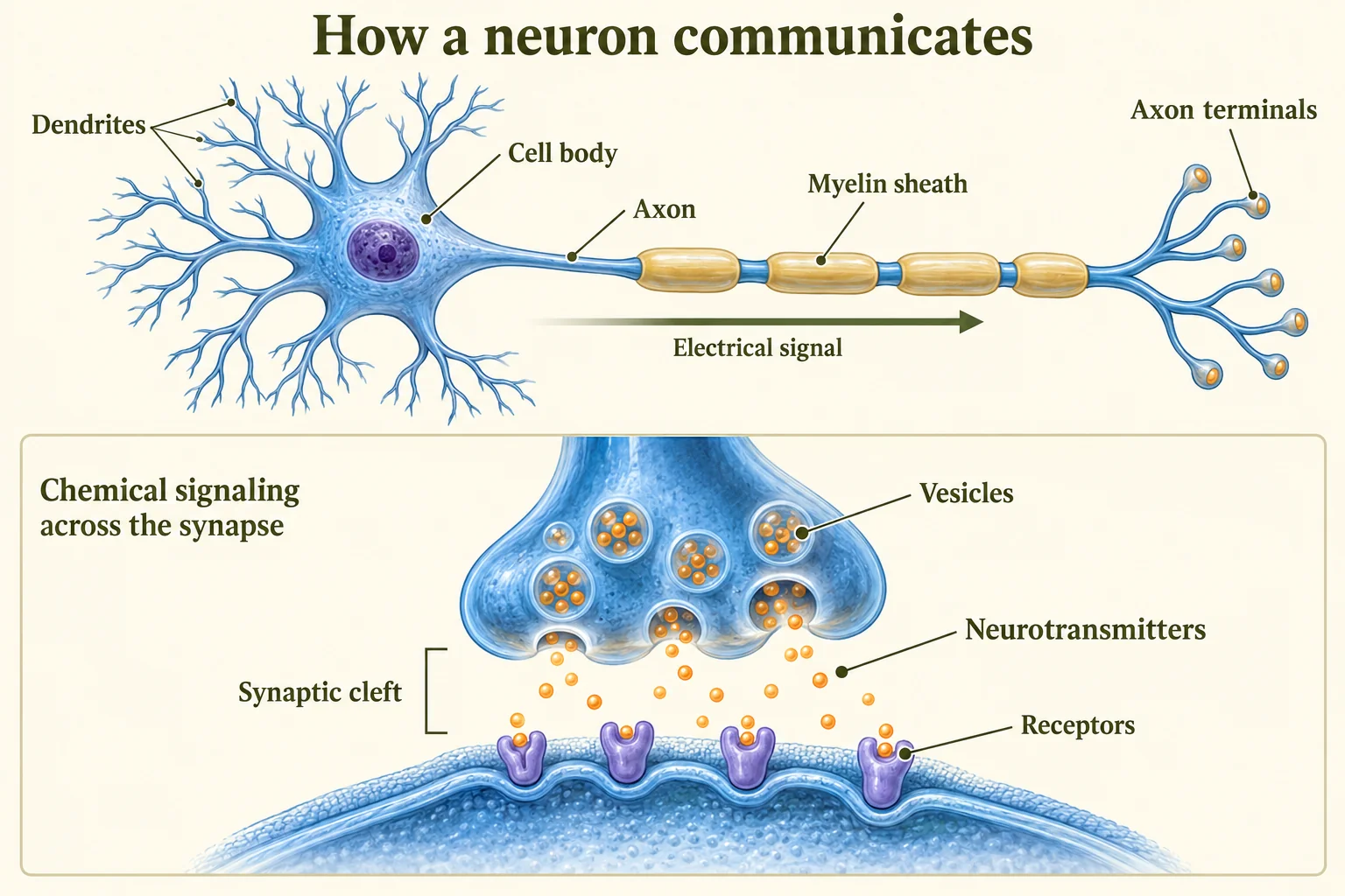
A typical neuron has specialized structures that enable it to receive, process, and transmit information efficiently.
1. Dendrites
Branch-like structures that extend from the cell body and receive incoming messages from other neurons.
Function: Act as receivers—gather chemical signals from neighboring neurons and convert them into electrical signals.
2. Cell Body (Soma)
The cell's life-support center containing the nucleus and organelles that maintain the neuron's health and processes incoming signals.
Function: Integrates signals from dendrites and determines whether the neuron will fire an action potential.
3. Axon
A long fiber that carries the electrical impulse (action potential) away from the cell body toward other neurons, muscles, or glands.
Function: Transmitter—conducts the action potential from the cell body to the axon terminals.
4. Myelin Sheath
A fatty coating produced by glial cells that wraps around the axon in segments, insulating it and dramatically speeding up neural transmission.
Function: Enables faster signal transmission through saltatory conduction (the signal "jumps" between gaps in the myelin).
Clinical Note: Loss of myelin leads to diseases like multiple sclerosis (MS), which disrupts neural signals and causes motor, sensory, and cognitive problems.
5. Terminal Buttons (Axon Terminals)
The ends of the axon that contain tiny sacs (vesicles) filled with neurotransmitters, which are released into the synapse when the action potential arrives.
Function: Release chemical messengers to communicate with the next neuron across the synaptic gap.
6. Synapse
A synapse is the communication junction between cells, including presynaptic and postsynaptic structures. At a chemical synapse, the synaptic cleft is the narrow extracellular gap across which neurotransmitters diffuse. Not all synapses are axon-to-dendrite or chemical.
Function: The communication junction where chemical transmission occurs.
Memory Aid: Neuron Signal Flow
Dendrites → Cell Body → Axon → Terminal Buttons → Synapse → Next Neuron
Think: RECEIVE (dendrites) → PROCESS (soma) → TRANSMIT (axon) → RELEASE (terminals)
Types of Neurons
The nervous system contains three main types of neurons, each with a specialized role in transmitting information.
1. Sensory Neurons (Afferent Neurons)
Carry incoming information from sensory receptors (eyes, ears, skin, muscles) to the central nervous system.
Example: Detect heat from a hot stove and send signals to the spinal cord and brain.
2. Motor Neurons (Efferent Neurons)
Carry outgoing commands from the central nervous system to muscles and glands, producing movement or secretions.
Example: Send signals from the spinal cord to hand muscles to pull away from danger.
3. Interneurons
Connect neurons within the brain and spinal cord, processing information between sensory and motor neurons.
Function: Enable complex processing, reflexes, and decision-making by relaying and integrating signals.
Spinal Reflex Arc
A spinal withdrawal reflex is a rapid, automatic response coordinated through spinal circuitry. It can begin without waiting for conscious awareness; sensory information also reaches the brain, and descending signals can modulate the response. Other reflexes can involve brainstem circuits.
Example: Touching a hot stove → pulling your hand away instantly
Reflex Arc Process:
- Sensory Neuron: Detects stimulus (heat on skin) and sends signal to spinal cord
- Interneuron: Processes signal in the spinal cord and relays it
- Motor Neuron: Sends command to muscle (pull hand away)
- Delayed Brain Awareness: You feel the pain only after reacting
Key Point: The reflex arc illustrates how the CNS and PNS work together to produce rapid, protective responses faster than conscious thought.
| Neuron Type | Function in Reflex Arc |
|---|---|
| Sensory Neuron | Detects stimulus (e.g., heat on skin) |
| Interneuron | Processes signal in spinal cord |
| Motor Neuron | Sends command to muscle (pull hand away) |
The Neural Firing Process
Neural firing is the process by which neurons generate and transmit electrical signals called action potentials.
1. Resting Potential
At the resting potential, a neuron maintains a membrane voltage commonly illustrated as , relative to the outside. It remains metabolically active and can receive changing input; the value varies by cell.
The neuron is polarized—ready to fire like a charged battery, with more negative ions inside the cell membrane and more positive ions outside.
State: The neuron is waiting for sufficient stimulation to trigger an action potential.
2. Threshold
Threshold is the membrane-voltage level at which an action potential is initiated, commonly illustrated as . It is not a universal fixed voltage or the same thing as stimulus intensity.
If incoming signals from other neurons push the neuron's internal charge to threshold or above, the neuron will fire.
If the stimulation is below threshold, the neuron does NOT fire and returns to resting potential.
3. Action Potential
The electrical impulse that travels rapidly down the axon when the neuron fires, transmitting information from the cell body to the axon terminals.
The action potential is also called "neural firing" or a "spike".
This is the fundamental unit of communication between neurons and represents the neuron's "message".
4. Depolarization
When threshold is reached, ion channels open and positive ions (sodium ions) rush into the axon, making the inside of the neuron less negative or even positive.
During the depolarizing phase of an action potential, membrane voltage can rise from an illustrative resting value of to a positive peak such as . Exact voltages vary; an action potential also includes repolarization and often an after-hyperpolarization.
Depolarization propagates down the axon like a wave, carrying the neural signal toward the terminal buttons.
5. All-or-None Principle
A neuron either fires completely or not at all—there is no partial firing.
Once the threshold is reached, the action potential has a fixed size and strength regardless of how strong the original stimulus was.
For a given neuron, stronger stimulation does not simply make each action potential taller. Intensity can be represented by firing rate and by recruitment of additional neurons, within biological limits.
Analogy: Trigger of a Gun
Pulling a trigger lightly does nothing. Pulling it hard enough fires the gun with the same force every time. The bullet's speed and force don't change based on how hard you pull—the gun either fires or it doesn't.
6. Refractory Period
The brief recovery time immediately after an action potential during which the neuron cannot fire again (or requires much stronger stimulation).
Types of Refractory Periods:
- Absolute Refractory Period: The neuron cannot fire under any circumstances
- Relative Refractory Period: The neuron can fire, but only with a stronger-than-normal stimulus
Purpose: Ensures the action potential travels in one direction and prevents signals from piling up. The neuron resets to resting potential during this time.
Additional Process: Repolarization
After depolarization, repolarization involves sodium-channel inactivation and increased outward potassium current. A brief after-hyperpolarization may occur. Ion gradients are maintained over time; repolarization is not simply sodium flowing back out through the same channels.
Worked voltage difference
This optional arithmetic example compares two membrane potentials; it is not a full action-potential equation.
The change is positive because the inside becomes less negative. Millivolts measure voltage; this calculation does not calculate firing frequency or signal speed.
Neural Transmission Process Summary
Step-by-Step Neural Firing
Step 1: Resting Potential
Neuron is polarized, ready to fire ()
Step 2: Threshold Reached
Enough stimulation triggers action (around )
Step 3: Action Potential
Electrical impulse travels down the axon
Step 4: Depolarization
Positive ions rush in, flipping the charge
Step 5: Refractory Period
Neuron resets, temporarily can't fire again
Step 6: Neurotransmitter Release
Chemical messengers cross the synapse to the next neuron
Synaptic Transmission
After the electrical action potential reaches the axon terminal, communication between neurons switches from electrical to chemical.
How Synaptic Transmission Works
- Action potential arrives at the axon terminal (terminal button)
- Neurotransmitters are released from vesicles into the synapse (synaptic cleft)
- Neurotransmitters cross the gap and bind to receptor sites on the receiving neuron's dendrites
- Binding causes changes in the receiving neuron (excitatory or inhibitory effects)
- Signal termination: Depending on the transmitter, reuptake into neurons or glia, enzymatic breakdown and diffusion help clear transmitter and limit signaling.
Key Point: The synapse is where neurons communicate chemically. Neurotransmitters are the chemical messengers that carry signals across this gap.
Reuptake
Reuptake is the process by which neurotransmitters are reabsorbed back into the presynaptic neuron (the sending neuron) after they have transmitted their signal.
This cleanup process stops the signal and prepares the neuron for the next transmission, preventing continuous stimulation.
Neurotransmitters: Chemical Messengers
Neurotransmitters are chemical messengers that carry signals across the synapse from one neuron to another.
Neurotransmitters can be excitatory (increase likelihood of firing) or inhibitory (decrease likelihood of firing).
AP Exam Alert
Only 8 neurotransmitters are tested on the AP Psychology exam. Focus on these and their primary functions.
| Neurotransmitter | Type | Primary Function |
|---|---|---|
| Dopamine | Depends on receptor and circuit | Reward, movement, attention, motivation (Parkinson's, schizophrenia) |
| Serotonin | Depends on receptor and circuit | Mood, sleep, appetite (depression) |
| Norepinephrine | Depends on receptor and circuit | Alertness, arousal, stress response |
| Glutamate | Excitatory | Learning and memory (most common excitatory neurotransmitter in CNS) |
| GABA (Gamma-Aminobutyric Acid) | Inhibitory | Calms neural activity, reduces anxiety (main inhibitory neurotransmitter) |
| Acetylcholine (ACh) | Depends on receptor and circuit | Muscle contractions, learning, memory (Alzheimer's disease) |
| Endorphins | Inhibitory | Natural painkillers, produce feelings of euphoria (released during exercise) |
| Substance P | Excitatory | Transmits pain signals |
Excitatory Neurotransmitters
Increase the likelihood that the postsynaptic neuron will reach threshold and fire an action potential (depolarize the membrane).
Think: "Go" signal for neural firing
Inhibitory Neurotransmitters
Decrease the likelihood that the postsynaptic neuron will fire by hyperpolarizing the membrane.
Think: "Slow down" signal for neural firing
Association is not a complete cause: Neurotransmitters have multiple functions and receptor-dependent effects. Parkinson’s disease involves loss of dopamine-producing neurons in particular motor circuits; schizophrenia and depression cannot be reduced to a single whole-brain transmitter level. A drug’s receptor action is different from its full clinical effect.
Hormones and Behavior
Hormones are chemical messengers similar to neurotransmitters, but they travel through the bloodstream rather than across synapses and act more slowly with longer-lasting effects.
While neurotransmitters work in milliseconds to seconds, hormones work over minutes to days and affect many organs and tissues.
| Hormone | Function |
|---|---|
| Adrenaline (Epinephrine) | Triggers fight-or-flight response; increases heart rate and energy |
| Leptin | Regulates appetite and energy balance; signals satiety |
| Ghrelin | Stimulates hunger |
| Melatonin | Regulates sleep-wake cycles (circadian rhythms) |
| Oxytocin | Roles in childbirth, milk release and social behavior; social effects depend on context, not a universal trust or “love” switch |
AP Exam Note
Scope: Focus on the listed hormones and their behavioral roles. Detailed endocrine-gland information is outside this topic’s current AP scope except the pituitary gland, which is included with brain structures.
Psychoactive Drugs and Neural Communication
Psychoactive drugs alter brain chemistry by affecting neurotransmitter systems, changing how neurons communicate and influencing behavior and mental processes.
How Drugs Affect Neurotransmitters
Drugs affect neurotransmitters in three main ways:
1. Agonist
Mimics or enhances neurotransmitter activity by binding to receptors and activating them or increasing neurotransmitter release.
Example: Heroin mimics endorphins and activates their receptors, producing euphoria and pain relief.
2. Antagonist
An antagonist blocks receptor activation by a transmitter or agonist. Blocking an excitatory pathway can reduce an effect; blocking an inhibitory pathway can increase downstream activity. An antagonist does not universally prevent neural firing.
Example: Caffeine blocks adenosine receptors, preventing the "calm down" signal and increasing alertness.
3. Reuptake Inhibitor
Blocks reabsorption of neurotransmitters back into the presynaptic neuron, increasing the amount available in the synapse.
Example: Prozac (SSRI) blocks serotonin reuptake, increasing serotonin levels to improve mood.
Memory Aid: Key and Lock Analogy
- Agonist: Like a duplicate key—fits and opens the lock (activates the receptor)
- Antagonist: Like a broken key—fits the lock but jams it so nothing can happen (blocks the receptor)
- Reuptake Inhibitor: Like keeping the original key in the lock longer (prevents cleanup)
Drug Class Effects
| Drug Class | Effect on Nervous System | Examples |
|---|---|---|
| Stimulants | Increase neural activity and arousal | Caffeine, nicotine, cocaine, amphetamines |
| Depressants | Decrease neural activity and slow body functions | Alcohol, benzodiazepines (Valium, Xanax) |
| Hallucinogens / perceptual-altering drugs | Distort perception and cognition | LSD and psilocybin; cannabis has distinct and overlapping effects |
| Opioids | Relieve pain and induce euphoria | Morphine, heroin, OxyContin |
Tolerance, Addiction, and Withdrawal
Tolerance
As the brain adapts to repeated drug exposure, more of the drug is needed to achieve the same effect.
Addiction
Characterized by compulsive drug-seeking despite harmful consequences, with disruption to natural reward systems (especially dopamine pathways).
Withdrawal
Physical and psychological distress when drug use stops, including:
- Physical symptoms: Shaking, sweating, nausea, pain
- Psychological symptoms: Anxiety, depression, intense cravings
Neural Transmission Disorders
Multiple Sclerosis (MS)
The immune system damages myelin (the insulating sheath on axons), disrupting action potential transmission.
Result: Slower or failed neural signals causing numbness, weakness, vision problems, and cognitive difficulties.
Myasthenia Gravis
The immune system blocks acetylcholine receptors, preventing neurotransmitter binding.
Result: Weakened muscle control, especially in eyes, face, and limbs.
AP Exam Strategy
Multiple Choice Tips
- Know neuron parts and their functions (dendrites, soma, axon, myelin, terminals)
- Understand the firing process: resting potential → threshold → action potential → refractory period
- Master the all-or-none principle: neurons fire completely or not at all
- Memorize all 8 neurotransmitters and their primary functions
- Distinguish between excitatory and inhibitory neurotransmitters
- Know drug mechanisms: agonist, antagonist, reuptake inhibitor
Free Response Question (FRQ) Tips
- Use precise terminology: action potential, threshold, depolarization, reuptake, synapse
- Explain processes step-by-step: trace neural firing from dendrites to axon terminals
- Connect neurotransmitters to behaviors: dopamine and reward, serotonin and mood, GABA and anxiety
- Apply drug effects: explain how agonists/antagonists change neural communication
- Use examples: reflex arc, MS disrupting myelin, SSRIs blocking serotonin reuptake
Quick Review Summary
The Big Picture
Neurons are the basic units of the nervous system that transmit information through electrical signals (action potentials) down the axon and chemical signals (neurotransmitters) across the synapse. Neural firing follows the all-or-none principle, and neurotransmitters can be excitatory or inhibitory. Psychoactive drugs alter behavior by acting as agonists, antagonists, or reuptake inhibitors.
Essential Concepts
- Neurons vs. glial cells
- Neuron structure (dendrites, soma, axon)
- Myelin sheath function
- Resting potential ()
- Threshold and action potential
- All-or-none principle
- Depolarization process
- Refractory period
- Synapse and neurotransmitters
- Excitatory vs. inhibitory
- 8 neurotransmitters (exam focus)
- Agonist, antagonist, reuptake
- Drug classes (stimulants, depressants)
- Tolerance, addiction, withdrawal
The Neuron and Neural Firing FAQs
Does a stronger stimulus make each action potential bigger?
For a given axon, action potentials are all-or-none. Intensity can be represented by firing rate and recruitment of neurons, rather than a larger spike.
Is a synapse the same as the synaptic cleft?
The synapse is the communication junction. The cleft is the narrow gap at a chemical synapse; presynaptic and postsynaptic components are also part of the junction.