
NUM8ERS · BIOLOGY STUDY NOTES
How Bacteriophages Kill Bacteria | Phages Explained
How can a virus kill a bacterial cell? Follow genome entry, phage production and cell lysis, then explore lysogeny, bacterial defences and the evidence behind phage therapy.
Watch the lesson ↓ Explore the notes ↓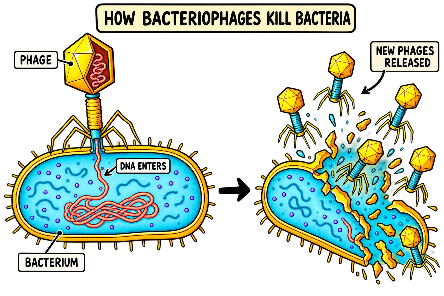
WATCH AND LEARN
How Bacteriophages Kill Bacteria
Watch the explanation, then use the notes below to study each step. Watch on YouTube ↗
1. What is a bacteriophage? Start with the parts
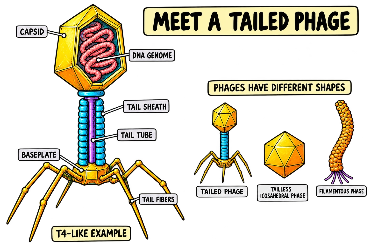
A bacteriophage, usually shortened to phage, is a virus that infects bacteria. A bacterium is a cell: it contains a membrane, genetic material, ribosomes and the machinery needed to carry out metabolism. A phage particle is a much smaller package of genetic instructions protected by proteins. It depends on a suitable bacterial cell to produce more phages. The word does not mean that the virus has a mouth or swallows the bacterium. [1]
The familiar picture looks like a tiny spacecraft with a head, a tail and several landing legs. That is a useful example of a tailed phage, not a portrait of every bacterial virus. Some phages have short tails, others have long flexible tails, and some are filamentous or lack a tail. Their genomes also differ. Many well-studied tailed phages contain double-stranded DNA, but phages as a group include other genome types. A shape in a diagram cannot tell you everything about a virus’s biology. [2]
Read the structure by asking what each part does
The capsid is the protein shell around the genome. Its job is to protect that genome between infections and help package it into a particle that can reach another host. In a T4-like example, the tail includes a tube surrounded by a contractile sheath. A baseplate and receptor-binding structures connect recognition at the bacterial surface with the delivery apparatus. The tail fibres are protein structures, not legs that walk or muscles that pull the virus around. [3]
Think of the drawing as a relationship between information, protection and delivery. DNA carries instructions. The capsid protects the instructions. Surface-recognition and tail structures help those instructions reach an appropriate cell. This is a better explanation than memorising a labelled shape without connecting each label to a function.
The colours in these diagrams help you follow components; bacteria and phages do not naturally have these classroom colours. The enlarged cutaways also make membranes, DNA and tail structures visible together, so their relative sizes should not be treated as measurements.
Is a phage alive?
This question depends on the definition of life being used. Phages have genomes and evolve, but a free phage particle does not independently grow, make proteins or reproduce as a cell does. For a biology answer, state the relevant property rather than relying on a single word: phages are acellular viruses that reproduce through suitable bacterial hosts. That sentence explains the dependency directly.
Check your understanding: If a diagram shows a phage with a contractile sheath, can you conclude that every phage has one? No. You can identify the illustrated example, but you need evidence before extending its features to the whole group. Keep that distinction in mind throughout this guide: we will use clear examples while pointing out where mechanisms vary.
2. How a phage finds a host and delivers its genome
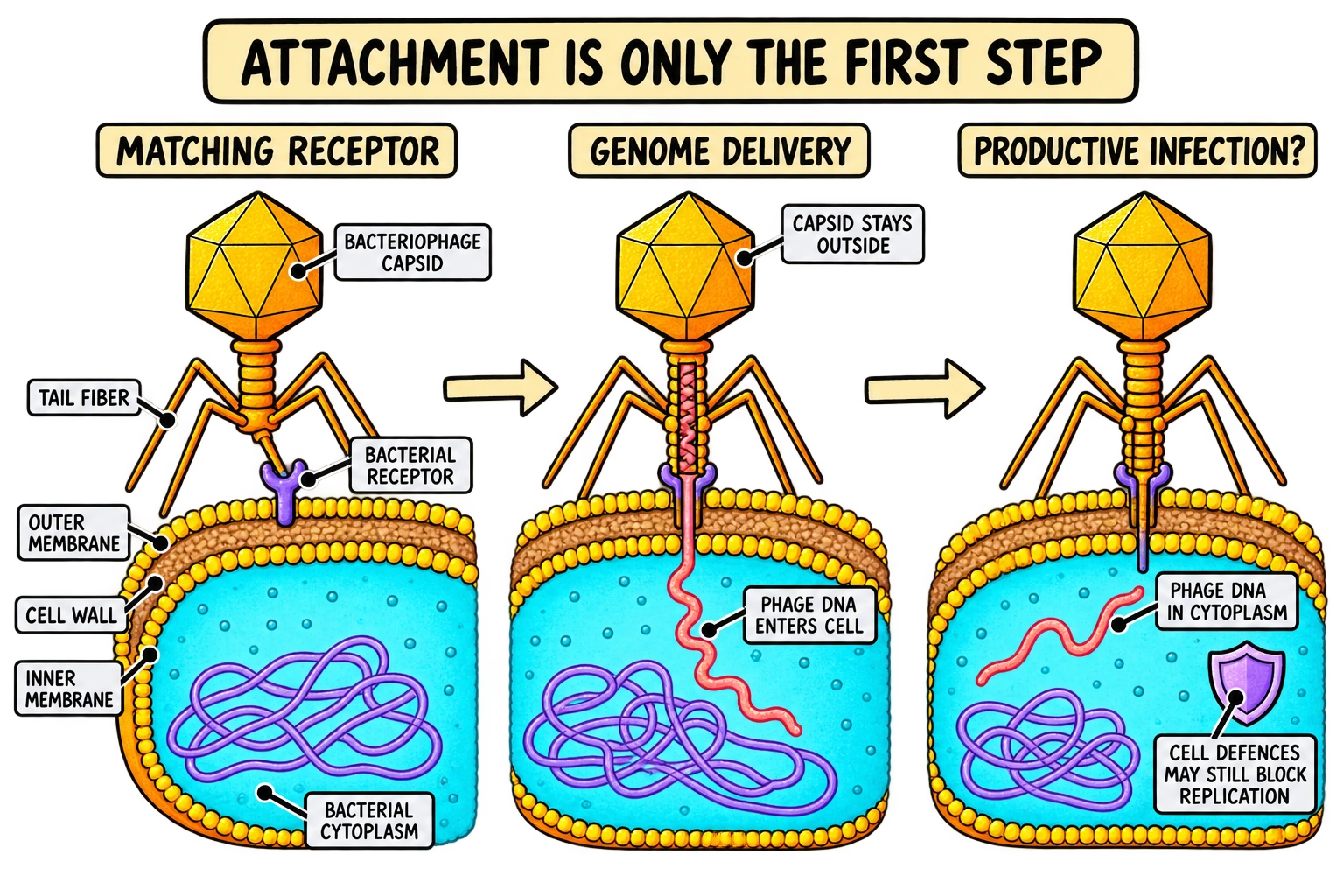
An encounter with a bacterium is only the beginning. To establish an infection, a phage must recognise a usable surface feature, deliver its genome and overcome whatever barriers operate inside that cell. These are separate checkpoints. A phage may succeed at the first and fail at the next. This is why host range means more than a simple list of bacterial names.
Recognition depends on molecular compatibility
A bacterial receptor is a surface molecule or structure that a phage can use during attachment. Receptors can include proteins, surface carbohydrates or appendages, depending on the phage. Receptor-binding proteins on the virus interact with these features. The comparison to a key and a lock is helpful if you remember its limit: real recognition can involve several contacts and structural changes, rather than one perfectly rigid key fitting one hole.
Phage lambda provides a specific example. Its tail interacts with the Escherichia coli outer-membrane protein LamB. A 2024 structural study examined how this receptor interaction is connected with the phage’s infection apparatus. The useful lesson is that attachment is a physical interaction between particular molecules, not a virus recognising the written name of a bacterial species. [4]
Different strains of the same bacterial species can have different surface features. Work on phage tailspike proteins has connected their sequence and structural variation with recognition of bacterial polysaccharides. Consequently, saying that a phage infects “E. coli” does not automatically mean it infects every E. coli strain. Equally, some phages infect a broader range than others. Specificity must be established for the phage–host combination being discussed. [5]
Genome entry is not whole-particle entry
For the tailed DNA-phage examples in the diagrams, the capsid stays outside while the genome passes through a delivery route into the bacterial cytoplasm. In a contractile-tailed example, changes in the baseplate and sheath help bring the tail tube into position. Other tailed phages use different entry machinery; not every phage has a sheath that contracts. The important shared question is how a protected genome crosses the host envelope. [6]
Once inside, the genome still needs a productive environment. The host might destroy the incoming nucleic acid, block expression of viral genes or stop the infection through another defence. Therefore attachment, genome delivery and productive infection are related outcomes, but they are not interchangeable observations.
Use this idea in an exam question
Suppose a phage attaches to two bacterial strains, but new infectious particles appear from only one. The observation does not require the phage to have attached incorrectly to the other strain. A later step could have failed. A good answer names a plausible checkpoint and then explains what additional evidence would distinguish it: was the genome delivered, copied, expressed or packaged?
Check your understanding: “The phage bound to the receptor, so the bacterium must lyse.” What is missing? Evidence that the infection progressed through genome expression, production of progeny and the relevant release pathway. Binding is evidence of contact, not proof that the entire cycle succeeded.
3. The lytic cycle: how one infection produces new phages
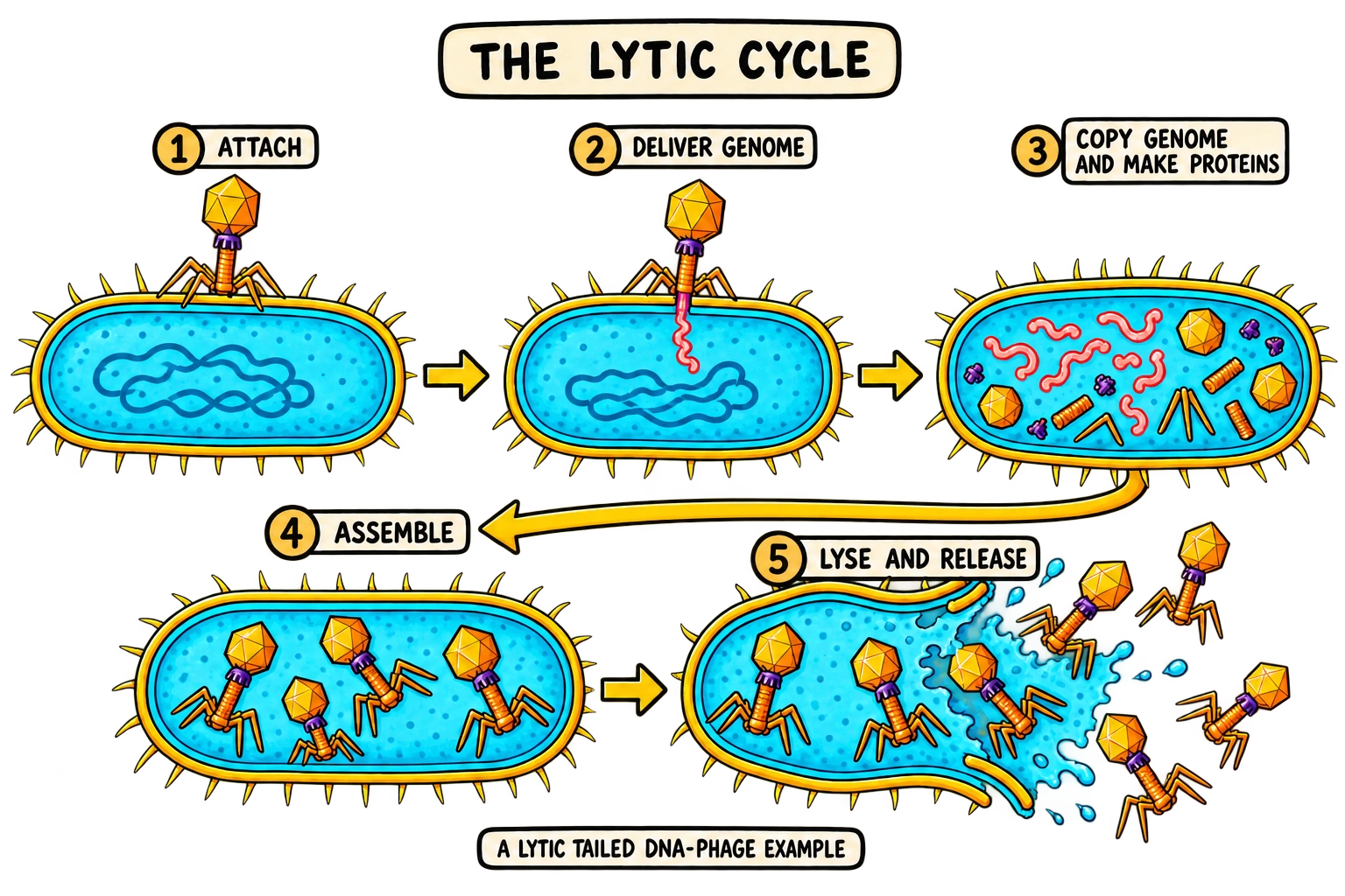
The lytic cycle is a productive infection that ends with rupture of the host cell and release of progeny phages. The word lysis refers to that breaking-open event. The complete story includes much more than the final rupture: the phage first needs access to the cell, then new genetic material and proteins must be made, and these components must become infectious particles.
For revision, learn five connected stages: attachment, genome delivery, biosynthesis, assembly and release. Different courses may call delivery “penetration” and assembly “maturation”. These labels describe overlapping parts of the same process. Explain the events behind the words rather than treating vocabulary differences as different life cycles. [1]
Biosynthesis means making the components
After successful entry, viral genes are expressed using a combination of host resources and phage-encoded functions. The host’s ribosomes translate suitable messenger RNAs into proteins. Nucleotides, amino acids and energy are required. The phage genome is copied, and proteins needed for replication, construction and eventual release are produced.
The contribution of the host and the virus differs between systems. For example, phage T7 uses the host RNA polymerase for early transcription and subsequently uses a phage-encoded RNA polymerase for other viral transcription. That example prevents a common overstatement: a phage does not necessarily depend on the host for every enzyme, even though it depends on a host cell for reproduction. [7]
Genome replication and protein synthesis must also be distinguished. Replication produces more genetic material. Transcription produces RNA from a DNA template in these DNA-phage examples. Translation produces proteins from messenger RNA. A capsid protein does not become DNA, and DNA copies do not automatically fold themselves into a complete virus.
The order matters because later steps need earlier products
An infection can fail even when some viral DNA has been copied. If structural proteins are missing, mature particles may not form. If particles assemble but remain trapped inside an intact cell, they cannot spread in the usual way. If released particles cannot recognise another suitable host, release does not guarantee another successful infection.
This gives you a useful reasoning chain: delivery provides instructions; gene expression and replication supply parts; assembly makes particles; release makes those particles available to encounter new hosts. Each arrow represents a dependency. In a short answer, naming the stages earns less explanatory value than showing what the next stage requires.
Do all lytic infections take the same time?
No fixed number of minutes applies to every phage and every condition. The host’s physiological state, the particular phage, temperature and other environmental factors can change infection dynamics. Similarly, the number of progeny released is variable. A textbook value for one well-studied system is not a universal phage constant. Quantitative work on phage growth established why timing and production need to be measured for the system being studied. [8]
Check your understanding: A cell contains phage DNA and capsid proteins. Has it necessarily produced infectious progeny? No. You still need evidence that genomes were packaged into correctly assembled particles that can initiate another productive infection.
4. Assembly and genome packaging: building an infectious particle
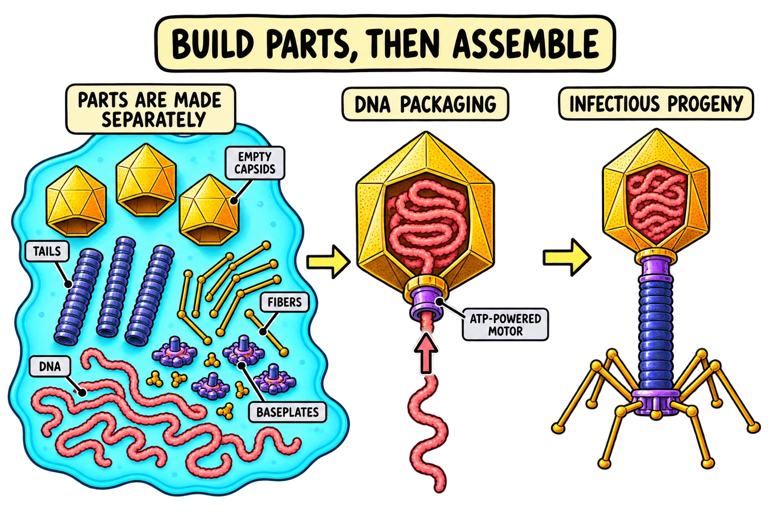
Making components is not the same as building a working structure. A pile of capsid proteins, tail proteins and copied DNA is not yet an infectious phage. Assembly arranges the components into the appropriate architecture. Maturation includes changes that make the assembled structure ready for its next biological task.
In T4, head formation, tail formation and long-tail-fibre formation can be described as separate construction pathways that ultimately join. This is different from bacterial reproduction. A bacterium can grow and divide into daughter cells; a phage population is generated through component production and particle assembly within a host. One incoming phage does not simply swell up and split into two identical tiny phages. [3]
DNA must go inside the right container
For many tailed double-stranded DNA phages, a packaging motor helps move the genome into a preformed head through a portal. The portal is a specialised entry point in the capsid. The motor uses energy from ATP hydrolysis; it is not a hand pulling on a DNA thread. The resulting DNA-filled head then needs the correct connections and closures to retain the genome and function during the next infection.
Experiments on the T4 packaging machine have investigated DNA movement into phage heads, including measurements at the level of individual motors. Such work supports the idea that packaging is an active molecular process. It should not be confused with DNA replication: copying the genome creates the material; packaging places that material into a transportable particle. [9]
The finished particle must also be ready to deliver
Assembly is a sequence of structural transitions, not just a final inventory of parts. A 2026 cryo-electron microscopy study of the T4 portal–neck–tail complex examined how changes after genome packaging help position the genome in an infectious virion. For students, the important point is that the connection between a packed head and a delivery-ready tail is itself a regulated structural problem. Knowing that DNA fits inside a capsid does not explain how it remains there and later exits correctly. [10]
Avoid turning this into a rule that every phage builds itself exactly like T4. Filamentous phages use a different architecture and assembly route. Their long, narrow particles demonstrate that the common need—protecting and transmitting a genome—can be met by different structures. [11]
A useful comparison: information versus infectivity
Imagine two samples in a classroom problem. One contains many copies of phage DNA but few functional particles. The other contains fewer DNA copies overall but many particles that successfully infect a test host. A DNA-detection result and an infectivity result could rank these samples differently. That would not automatically make either measurement wrong; they answer different questions.
When reading an experimental description, underline exactly what was measured: genome copies, protein abundance, visible particles or infectious units. Those quantities can be related, but they should not be substituted for one another without evidence.
Check your understanding: Why might a packaging defect reduce the number of infectious phages even if genome replication continues? The copied DNA has not been correctly incorporated into particles capable of protecting and delivering it to the next host.
5. How phages kill bacteria: breaking the cell envelope
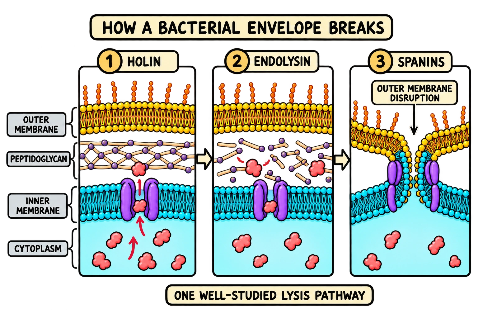
The usual cartoon ends with a bacterium bursting. To explain how bacteriophages kill bacteria, connect that picture to the structures that hold the cell together. The envelope is a physical barrier, and the cell wall provides mechanical support. Releasing progeny requires the phage to overcome the relevant barriers, not simply to produce enough particles to make the bacterium “too full”.
Follow the envelope from inside to outside
In a typical Gram-negative example, the inner membrane surrounds the cytoplasm. Outside it lies a thin layer of peptidoglycan, a mesh-like cell-wall material. An outer membrane forms an additional barrier. These layers have different chemical properties, so breaking one does not automatically remove the others.
Many double-stranded DNA phages use a coordinated lysis system. In the classical pathway, a holin acts at the inner membrane, an endolysin breaks down peptidoglycan, and spanins help disrupt the outer membrane of a Gram-negative host. This is an example of division of labour between proteins. The process is controlled in time rather than being a passive consequence of a cell filling with viruses. [12]
Holin: opening the inner-membrane barrier
In the canonical pathway, holin proteins accumulate in the cytoplasmic membrane and then form lesions that allow an endolysin to reach its cell-wall target. Imaging and biochemical work on lambda holin has examined the large assemblies associated with this membrane-permeabilising role. A holin is not the enzyme that digests the peptidoglycan mesh; its membrane function makes the enzyme’s action possible in this pathway. [13]
This distinction helps with mechanism questions. If an endolysin is present but cannot reach its substrate, simply detecting that enzyme does not prove that lysis should happen. Location matters as much as presence. A protein can be functional in principle yet separated from the structure on which it needs to act.
Endolysin: weakening the peptidoglycan wall
An endolysin is an enzyme that cleaves bonds in peptidoglycan. Once the wall loses the support it normally provides, the cell becomes vulnerable to its physical conditions. However, in the Gram-negative systems being described, the outer membrane can still present a barrier after wall degradation. The sequence therefore cannot be reduced to “one enzyme dissolves the whole bacterium”. [14]
Keep wall and membrane separate in your answer. Peptidoglycan is not a lipid bilayer. Saying that an endolysin “cuts the membrane” hides the specific target and makes it harder to explain why additional lysis proteins can be needed.
Spanins: dealing with the outer membrane
Experiments on lambda spanins support a mechanism in which they bring the inner and outer membranes together and promote membrane fusion after the peptidoglycan barrier has been removed. This provides a clear example of why the third envelope layer matters. The host can fail to release phages efficiently even when some earlier damage has occurred. [15]
Learn the example without making it universal
Not every bacterium has the same envelope, and not every phage uses identical release proteins. Gram-positive bacteria lack the typical Gram-negative outer membrane. Some phages use alternative holin/endolysin arrangements, and certain small phages disrupt cell-wall synthesis through other mechanisms. Filamentous phages can even leave a living cell without lysing it. [16] [17]
For a strong short answer, say: in this lytic phage–host system, phage proteins disrupt the envelope, allowing progeny to escape and killing the host cell. Then name the specific barriers and proteins if the question asks for detail. That wording gives both the mechanism and its appropriate scope.
Check your understanding: Why is “the phages explode the bacterium because there are too many of them” incomplete? It skips the phage-encoded lysis machinery and the different structural barriers that machinery must overcome.
6. Lysogeny: why an infected bacterium may stay alive
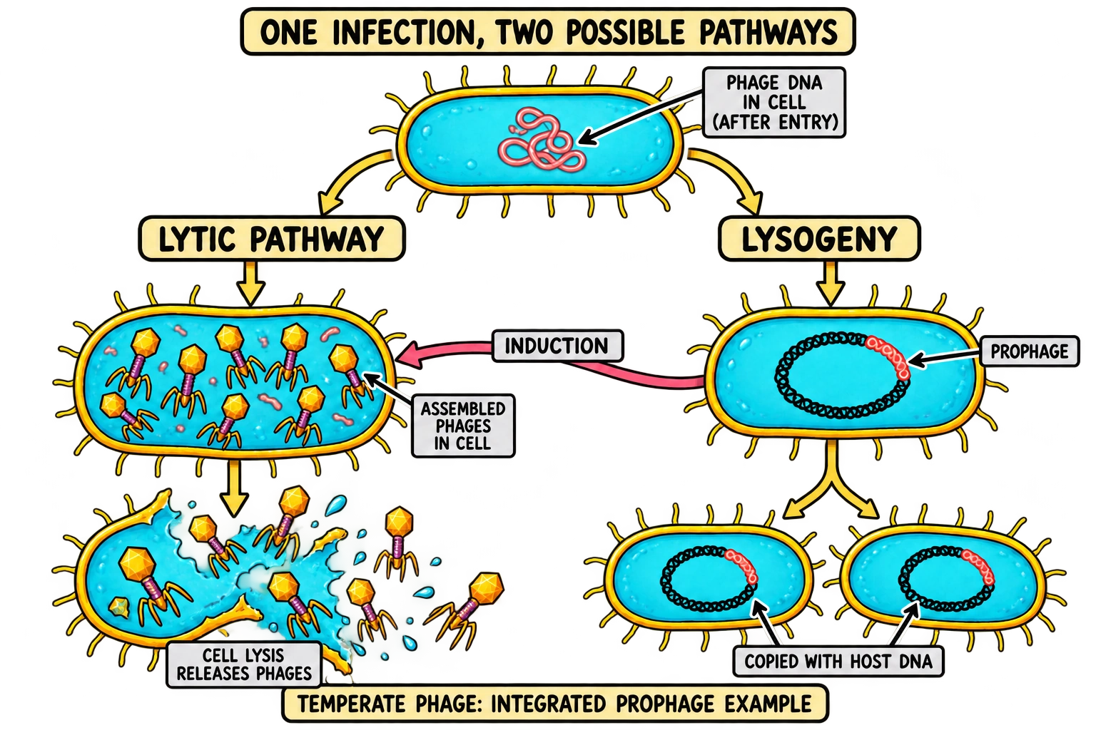
An infection does not always proceed immediately to lysis. Temperate phages can establish lysogeny, a state in which the viral genome is maintained in the host lineage instead of being used immediately for a destructive round of progeny production. Phage lambda is a classic example. A bacterium carrying the maintained phage genome is called a lysogen.
What is a prophage?
In the familiar lambda-like case, phage DNA integrates into the bacterial chromosome. This maintained viral genome is the prophage. When the chromosome is copied and the cell divides, the integrated DNA can be passed to daughter cells. The diagram colours the phage-derived segment separately so you can track it through the bacterial lineage. It does not show whole phage particles growing inside each daughter cell.
Integration is a useful example, but do not define every prophage solely by integration into a chromosome. Some temperate phages maintain their genomes in other stable forms. The broader point is maintenance of the viral genome in association with the host lineage, with productive development held under regulation.
A maintained genome is not necessarily silent in every respect
The phrase “the virus is asleep” is easy to remember but can be misleading. Lysogeny involves gene regulation. Some genes help maintain that state, and others can affect the host’s properties. In lambda, a regulatory switch involving the CI repressor helps keep the lytic programme suppressed. Research on the switch shows why the relationship is more precise than a particle simply waiting inside a cell. [18]
This is also why prophage and phage particle must not be confused. One is the maintained genetic state; the other is an assembled infectious package. A lysogen can carry phage information without containing a collection of mature virions ready to burst out at that moment.
Induction: a change of programme
Induction is the transition from a maintained prophage state towards productive development. In lambda, DNA-damage signalling can interfere with the regulatory system that maintains lysogeny. Researchers have also investigated how a bacterial product that damages DNA can stimulate prophage induction. Such findings connect the switch to the host’s biological environment. [19]
Induction is not a statement that every stressed bacterium must burst. The outcome depends on the prophage, the host and the relevant signal. Some prophage genomes are defective and cannot complete an infectious cycle. In a diagram showing a functional temperate phage, an induction arrow should lead from lysogeny towards phage production, rather than suggesting that all lytic infections must first pass through chromosome integration.
| Feature | Lytic pathway | Lysogeny |
|---|---|---|
| Main outcome | Progeny are produced and the host lyses in the example described | Phage genome is maintained in the host lineage |
| Genome state | Replicated for productive infection; integration is not a required first step | Often integrated as a prophage, but maintenance forms vary |
| Host division | Not the route used to release the lytic progeny | Can pass the maintained phage genome to daughter cells |
| Host fate | Lysis kills the infected cell | No immediate lytic destruction is required |
| Possible transition | A virulent phage need not establish lysogeny | Induction can lead a functional temperate phage towards productive development |
Do not confuse lysogeny with chronic release
These are different ideas. In lysogeny, the phage genome is maintained while destructive productive development is repressed. In a chronic filamentous infection, new particles can be assembled and extruded from a living bacterium. Direct imaging of filamentous phages emerging from living cells demonstrates that release and immediate host lysis are not inseparable events. [17]
Check your understanding: A phage-derived DNA segment is inherited by daughter bacteria, but no new infectious particles are detected. Which idea fits the observation? Maintenance in a lysogenic state is plausible. The evidence does not describe the final lytic-release stage merely because viral DNA is present.
7. Gene transfer and lysogenic conversion: a different kind of impact
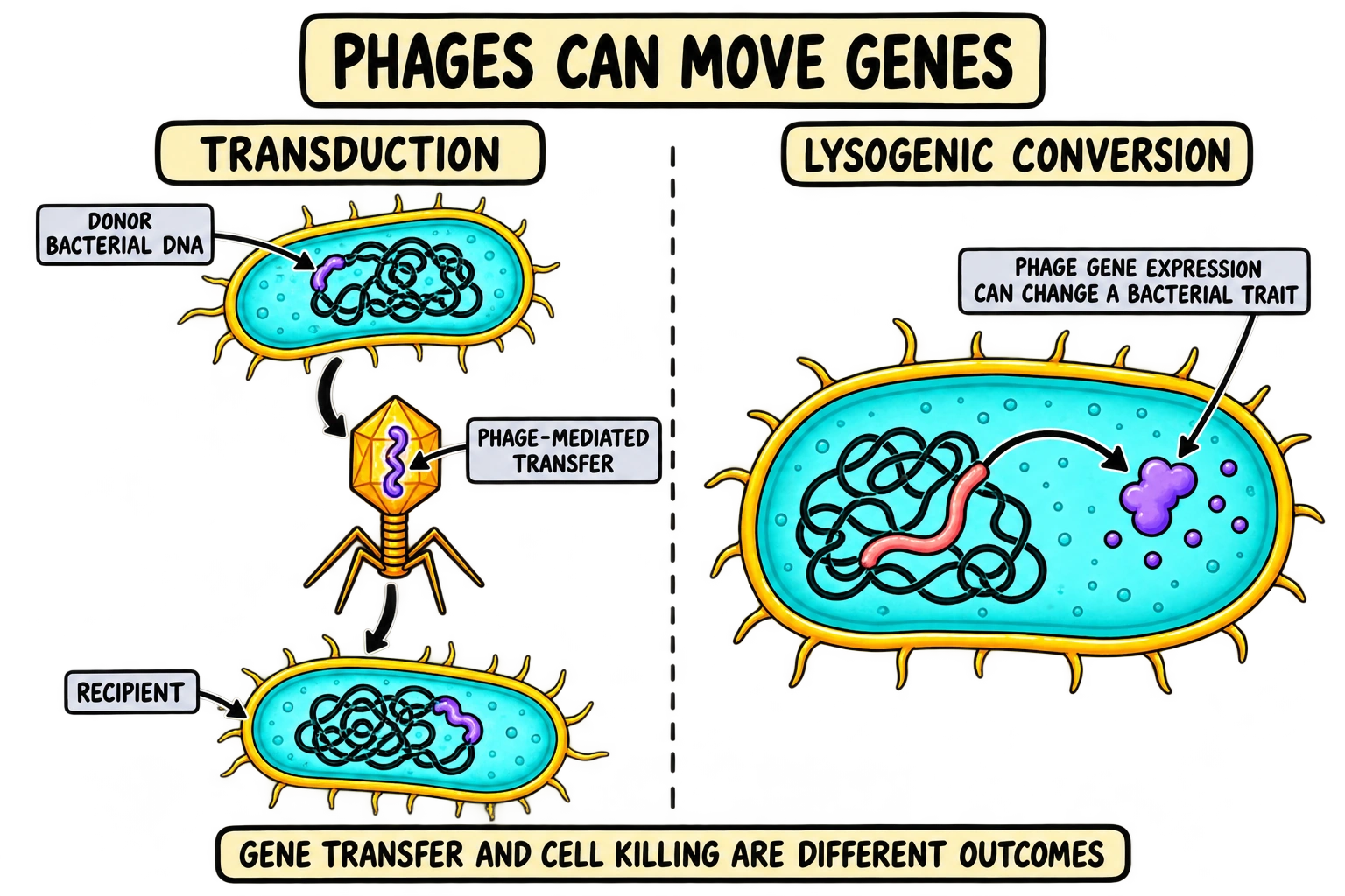
Phages can change bacterial populations by moving genetic information as well as by killing cells. Two terms help you keep these effects separate: transduction describes phage-mediated transfer of bacterial DNA, while lysogenic conversion describes a change in a host trait caused by genes associated with a prophage. Neither term simply means “the cell burst”.
Transduction moves bacterial DNA between cells
During some phage-associated processes, bacterial DNA can be carried in a particle and delivered to another bacterium. If that DNA is retained and expressed appropriately, it may contribute to a heritable change. Zinder and Lederberg’s work on genetic exchange in Salmonella helped establish phage-mediated transfer as a major concept in bacterial genetics. [20]
In generalized transduction, fragments from different regions of the bacterial chromosome can potentially be transferred through packaging errors in suitable phage systems. In specialized transduction, transfer is associated with particular bacterial sequences near a prophage integration site when an abnormal excision event includes neighbouring DNA. The words “generalized” and “specialized” refer to the kinds of bacterial DNA that can be moved, not to whether the virus is generally useful or highly dangerous. [21]
Transfer is not automatically successful. DNA must arrive in a recipient, avoid destruction and become stably maintained if it is to contribute to an inherited trait. A particle carrying bacterial DNA may also lack the complete viral genome needed to produce another generation of phages. Keep delivery of a DNA fragment separate from productive phage reproduction.
Lysogenic conversion changes a bacterial trait
A prophage can carry genes that alter the host’s phenotype. A well-established example concerns cholera toxin genes carried by the filamentous phage CTXφ. Waldor and Mekalanos demonstrated the phage association of those genes in Vibrio cholerae. This example shows that a virus of bacteria can affect human disease indirectly through the properties of its bacterial host. [22]
It does not mean that every prophage carries a toxin gene, or that every phage infection makes a bacterium more harmful. The correct statement is conditional: some phage-associated genes can alter bacterial traits, and the effect depends on the genes and the host. The same care applies to discussions of antibiotic-resistance genes; not every detected gene is functional or efficiently transferred.
Use the terms to compare outcomes
Imagine three observations. In the first, a cell releases infectious phages and lyses. In the second, a recipient bacterium acquires DNA from another bacterial strain through a phage-associated transfer event. In the third, a lysogen expresses a trait encoded by its prophage. The relevant explanations are lytic reproduction, transduction and lysogenic conversion respectively. These outcomes may be connected biologically, but naming one does not explain the others.
This distinction is especially useful when you read the word “harmful”. A phage can harm a bacterial cell by lysing it, yet some phage genes can increase the harm that a bacterial population causes to another organism. Always ask harmful to which organism, through which mechanism? That question turns an ambiguous statement into a testable biological claim.
Check your understanding: Does a toxin-associated prophage imply that humans are the phage’s reproductive host? No. The phage infects bacteria; a bacterial product can then affect humans. Direct viral host range and an indirect disease effect are different relationships.
8. Bacterial resistance: why phages do not always win
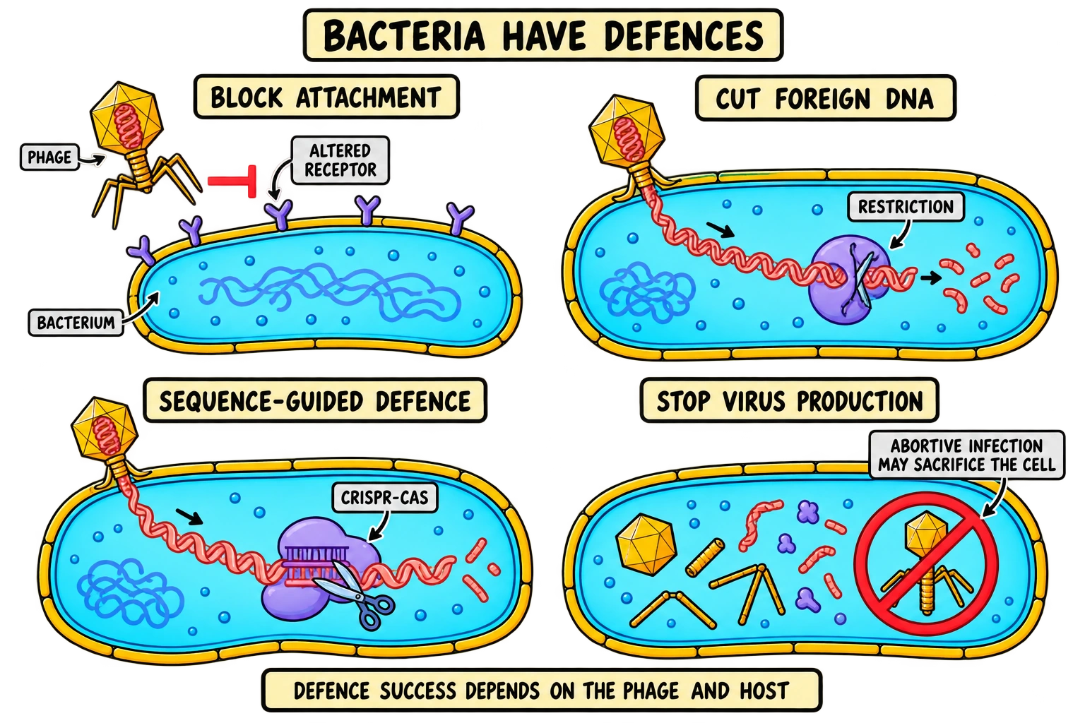
Bacteria are not passive containers. Their surfaces and intracellular systems can prevent or interrupt phage infection. At the same time, phages evolve ways around some barriers. The result is a continuing set of evolutionary interactions rather than a guaranteed victory for either side.
Block recognition or genome delivery
A change in a bacterial surface receptor can reduce attachment by a phage that depends on that receptor. Other surface changes can hide a receptor or make it less accessible. This helps explain why closely related bacterial strains can differ in susceptibility, and why a population may become less susceptible after phage exposure.
Resistance can carry a cost. A receptor might also help the bacterium transport nutrients or carry out another function. In research on phage OMKO1 and Pseudomonas aeruginosa, phage selection was associated with changes affecting an efflux-pump component and increased antibiotic sensitivity under the conditions studied. That is an interesting evolutionary trade-off, not proof that every phage-resistant bacterium becomes antibiotic-sensitive. [23]
Destroy or recognise incoming genetic material
Restriction–modification systems can discriminate between appropriately modified host DNA and some incoming DNA, allowing restriction enzymes to attack susceptible foreign material. CRISPR–Cas systems can provide sequence-guided defence. In a foundational 2007 study, changes in phage-derived spacer sequences changed bacterial resistance, supporting the role of CRISPR-associated sequence information in antiviral protection. [24]
The word “memory” is useful here only if it is tied to a physical mechanism. A bacterium does not remember a phage through thoughts or antibodies. Particular nucleic-acid sequences and proteins can enable recognition of matching targets. Not every bacterium has the same defence system, and a matching sequence does not make all infections impossible under every circumstance.
Stop the infection, even at a cost to the infected cell
Some defensive systems interrupt phage production by disrupting the infected cell’s own functions. Abortive infection can reduce phage spread even if the individual infected cell does not survive. That outcome is easier to understand when you distinguish the fate of one cell from the survival of neighbouring relatives.
The defensive repertoire extends far beyond the few systems introduced in school courses. Doron and colleagues experimentally validated previously unrecognised systems in 2018. A 2026 study used shared protein-domain patterns to identify and test additional defence arrangements. For revision, you do not need to memorise every system name. You do need to understand that bacterial immunity is diverse and that attachment alone cannot establish the final outcome. [25] [26]
Selection is not purposeful mutation
Luria and Delbrück’s classic work investigated whether phage resistance could arise before exposure. Their results supported the existence of resistant variants produced independently of the later selection event. A phage can therefore increase the proportion of resistant bacteria by removing susceptible competitors, rather than instructing each bacterium to make exactly the mutation it needs. [27]
Phage counter-defences complicate the story further. Some phages inhibit CRISPR-associated processes or otherwise interfere with bacterial protection. Consequently, host susceptibility is a property of an interaction: a defence that blocks one phage might fail against another. [28]
Check your understanding: After phage exposure, a culture contains many resistant bacteria. Does that observation alone prove that exposure created the resistance mutation? No. Pre-existing resistant variants may have survived and multiplied. You need a suitable experimental design to distinguish explanations.
9. Phages in ecosystems and the microbiome
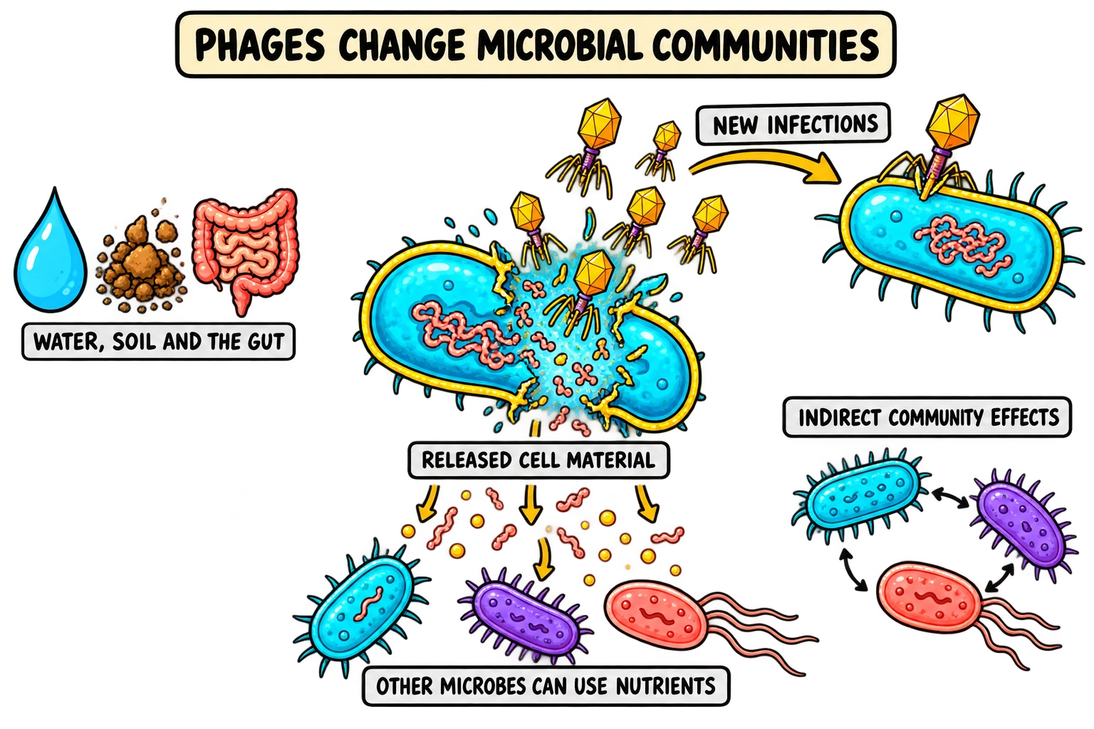
Phages occur wherever suitable bacterial hosts and environmental conditions allow them to persist. Water, soil and animal-associated microbial communities contain many different phage–host relationships. Their ecological importance comes from what those interactions do: removing susceptible cells, releasing cell contents, changing competition and moving genes.
Lysis redistributes material
When a bacterial cell lyses, some material that was contained in a cell becomes available in other forms. Other microbes may use released organic molecules and nutrients. In aquatic ecosystems, this can redirect material through microbial processes rather than leaving it in an intact bacterial cell that might have been consumed by a larger organism.
Measurements in the Southern Ocean have examined how lytic and lysogenic viral infection varies between locations and contributes to biogeochemical cycling. Such studies are important because a single universal percentage of “bacteria killed by viruses” would hide large differences between ecosystems and conditions. For an explanation, follow the material and the affected organisms before quoting an abundance estimate. [29]
The term viral shunt is often used for the redirection of cellular material into dissolved or particulate pools through viral lysis. It is a flow-of-matter idea, not a claim that every released molecule takes the same path. Environmental measurements are needed to establish the scale and destination of the flow in a particular system.
A specific target can still have indirect effects
Imagine a community in which bacterium A competes with bacterium B for a nutrient. A phage that suppresses A might indirectly change the growth of B, even if it cannot infect B. Alternatively, if B depends on a product made by A, suppressing A could reduce B’s growth. These are different predictions because competition and dependence connect the organisms differently.
A mouse-model study using a defined gut microbiota demonstrated both direct effects on susceptible bacteria and cascading effects on other community members, together with changes in the metabolome. The result supports a careful distinction: phage specificity at the infection step does not guarantee that all ecological consequences remain confined to one bacterial strain. It also does not establish the same response in every human gut. [30]
The mucus example: an interaction, not a universal shield
Research on mucus-associated phages showed that some phage–mucin interactions can enrich phages near mucosal surfaces. Experiments also examined protection of underlying cells from bacterial challenge. This led to the bacteriophage-adherence-to-mucus model, a way to investigate how viruses of bacteria can interact with animal-associated habitats. [31]
For students, its value is in the connected mechanism: surface association can change encounter rates between phages and bacteria, and those encounters can affect a larger biological system. It should not become the unsupported statement that “all phages in the gut protect us” or that a person should deliberately consume environmental phages.
Keep scale clear
An effect on one bacterial cell, a bacterial population, a microbial community and an animal host are different levels of explanation. Moving between them requires evidence. A microscope image of one lysed cell cannot establish a whole-ecosystem nutrient budget. A change in a mouse microbial community cannot, by itself, prove clinical benefit in humans.
Check your understanding: Can a phage alter the abundance of a bacterium it cannot infect? Yes, indirectly, through changes in interacting community members or resources. That explanation needs a proposed ecological link; it does not require an expansion of the phage’s direct host range.
10. How scientists study phages: plaques, growth and evidence
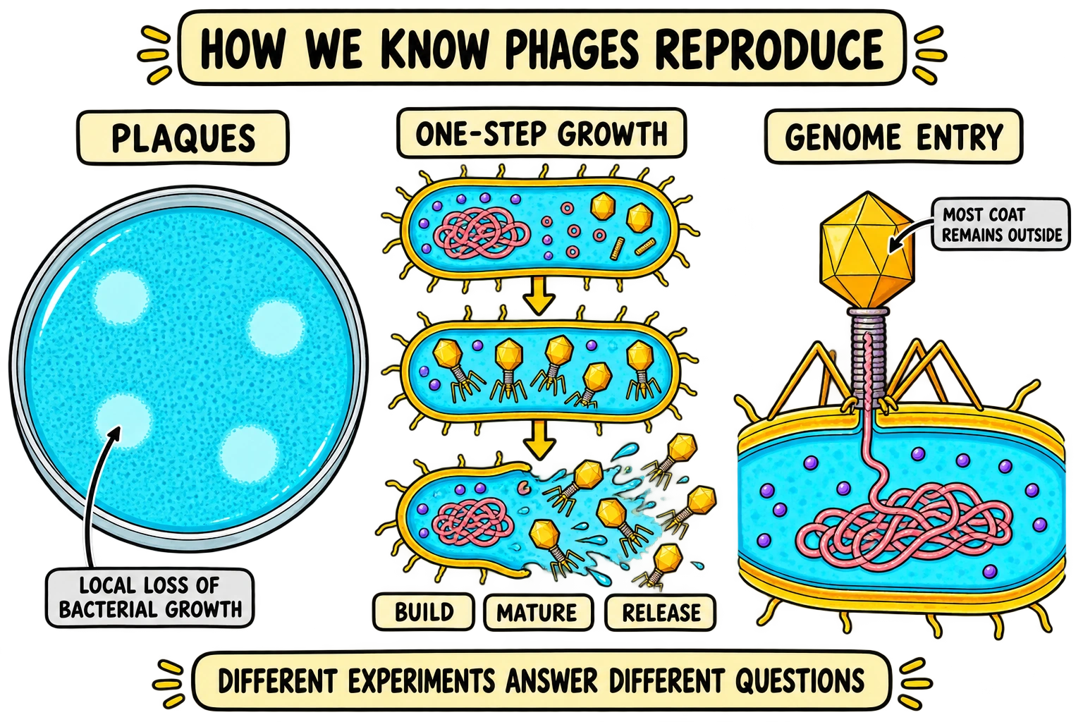
Different experiments reveal different parts of an infection. An image can show particle shape. A nucleic-acid test can detect genome material. An infectivity assay can show that material initiates productive infection under defined conditions. None of these observations should be asked to answer a question it was not designed to test.
What does a plaque show?
A plaque is a local region of reduced bacterial growth or clearing in a susceptible bacterial lawn associated with phage activity. It is not one enormous virus. Repeated infection and spread through nearby susceptible cells can produce a visible region even though the individual particles are too small to see unaided.
Plaque counting provides an operational measure called plaque-forming units, or PFU. A PFU is defined by what produces a plaque in the assay. It is therefore an infectivity measure for a particular host and set of conditions, rather than a direct count of every physical particle in a sample. The foundations of quantitative phage growth research included showing how plaque counts related to phage concentration. [8]
The one-step growth idea
In a suitably controlled population of infected cells, researchers can follow the appearance of infectious progeny over time. The latent period concerns the interval before extracellular progeny are released in the defined experiment. The eclipse period concerns the earlier interval during which mature infectious particles cannot yet be detected inside infected cells using the appropriate analysis. The endpoints differ because mature particles can appear before the cell releases them. [32]
Do not label every flat segment of a graph “nothing is happening”. Genome replication, gene expression and assembly may be occurring while extracellular infectivity remains low. A measurement can remain flat because the measured material is not yet outside the cell, not because all molecular activity has stopped.
Worked example: average burst size
Practice example—not experimental results: Assume a question gives 300,000 productively infected cells and a corrected yield of 24,000,000 newly released PFU. Also assume that residual input phages and later infection rounds have already been excluded. The average infectious yield per infected cell is:
The answer is 80 PFU per infected cell under those assumptions. It is an average, not proof that each cell released exactly 80 particles. It is also not automatically a physical-particle count: infectious yield and total particles can differ. State what is in the numerator and denominator before calculating.
Worked example: correcting a plaque count for dilution
Practice example—not a laboratory protocol: A question reports 40 plaques from 0.1 mL of a sample diluted to one millionth of its original concentration. To recover the original sample’s titer, account for both the tested volume and the dilution fraction:
The result describes PFU per millilitre for the assay described. It does not directly tell you whether a treatment works in a patient, whether every counted particle contains the same genome, or how many bacteria were present in the original environment. A correct numerical answer should retain the meaning of the unit.
What the Hershey–Chase experiment contributed
Hershey and Chase used differently labelled components of phage T2 to investigate the roles of DNA and protein during infection. Their observations supported the importance of the DNA that entered the bacterial cells, while much of the labelled coat material remained outside. The appropriate conclusion concerns that phage system and the experimental evidence; it is not that every virus in nature has DNA rather than RNA. [33]
Think like a scientist: A positive DNA test and a low PFU count are not necessarily contradictory. The first can include genetic material that does not correspond to a functional infectious particle. Ask what each method detects, then compare the results on that basis.
11. Phage therapy: what the evidence supports
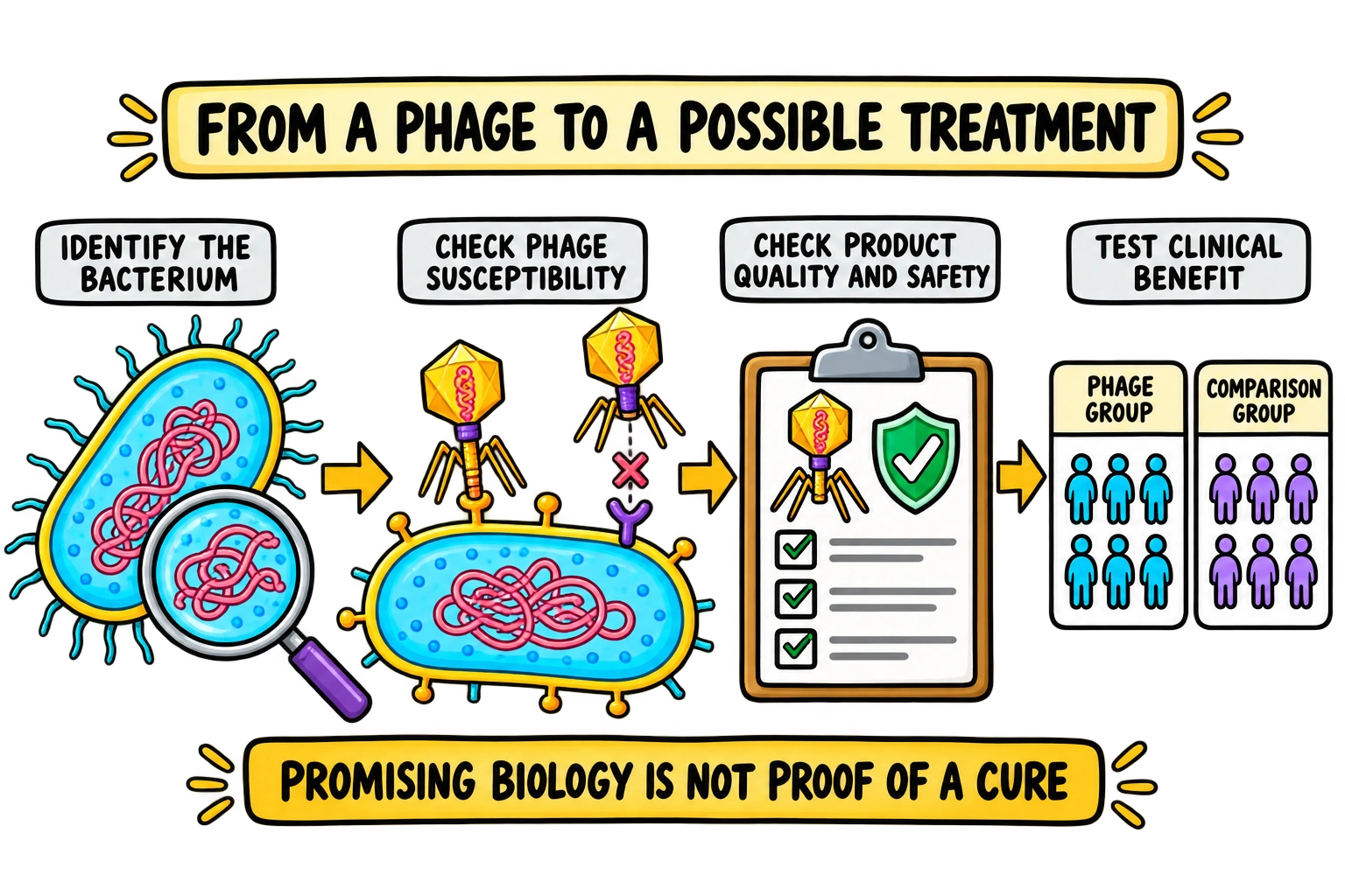
Phage therapy investigates or uses bacteriophages to target bacterial infections. Its biological appeal is clear: a suitable lytic phage can infect a susceptible bacterium through mechanisms different from those targeted by many antibiotics. However, a plausible mechanism, a result in a dish and a demonstrated clinical benefit are separate levels of evidence.
The World Health Organization’s 2025 report on building evidence for phage therapy highlights the need to develop and evaluate this approach in the broader response to antimicrobial resistance. That is a research and public-health opportunity, not a statement that any phage preparation is an established replacement for antibiotics. [34]
Why matching and product quality matter
A patient’s bacterial isolate must be susceptible to the phage or combination being considered. A phage cocktail contains multiple phages and may broaden coverage, but its components still need to be evaluated. Narrow infection specificity may help limit direct effects on unrelated bacteria, while community interactions can still create indirect effects.
Researchers also need to examine the identity and genetic properties of the phages, manufacturing consistency, purity and activity. Potentially unsuitable genes, bacterial contaminants and inflammatory material are relevant concerns. Reaching the infection site, retaining infectivity and interacting with the immune system add further challenges. Regulatory discussions by the transatlantic taskforce describe these issues as part of developing a defined biological medicinal product. [35]
Read case reports as case reports
In a widely discussed 2019 report, a patient with a disseminated drug-resistant Mycobacterium abscessus infection received a three-phage combination in a complex clinical setting. The report helped demonstrate the possibilities of carefully selected phages. It does not provide the same level of evidence as a large randomised comparison, and it should not be turned into a universal cure rate. [36]
When several treatments are given together, improvement cannot automatically be assigned entirely to one component. Clinical reasoning must consider the underlying disease, other therapies, timing and the outcomes measured. This is why a compelling individual story can motivate further research without settling every question about effectiveness.
A recent trial example, with the limits kept visible
A 2025 Nature Communications paper reported Part 1 of a randomised, double-blind, placebo-controlled study of nebulised BX004-A in nine adults with cystic fibrosis and chronic Pseudomonas aeruginosa infection. The study examined safety and tolerability and included exploratory bacterial-burden outcomes. Its small, early-stage design matters when interpreting the findings: it was not a definitive demonstration that phages cure all such infections. [37]
The paper also has a 2026 author correction concerning samples included in an analysis and associated figures/data. The correction states that the changes do not affect the study’s results or conclusions. Read the updated paper together with that correction. This is a practical example of checking the current scientific record rather than repeating an old summary without checking for updates. [38]
Promising does not mean universally effective or available
Phage resistance, difficult access to the infection site and differences between laboratory and patient conditions can all affect outcomes. Some combinations with antibiotics may be useful; others may behave differently. Evidence needs to be specific to the phage product, bacterial target, disease setting and comparison being studied.
In the United States, FDA discussions distinguish clinical trials from individual-patient expanded access to investigational phage products. Permission to investigate or provide access under such a pathway is different from demonstrating effectiveness or obtaining general marketing approval. Other jurisdictions have their own frameworks. [39]
For a student answer, the balanced conclusion is: phage therapy is a scientifically grounded approach being evaluated for particular bacterial infections, with important questions about matching, delivery, safety and clinical benefit. It belongs within qualified clinical and research systems, not home treatment with uncharacterised environmental material.
Check your understanding: Why is “the phage killed bacteria in a dish” insufficient proof of a cure? It does not establish delivery to the infected tissue, the response of the patient, comparative clinical benefit or the safety of the finished preparation.
12. Revision notes, worked reasoning and common questions
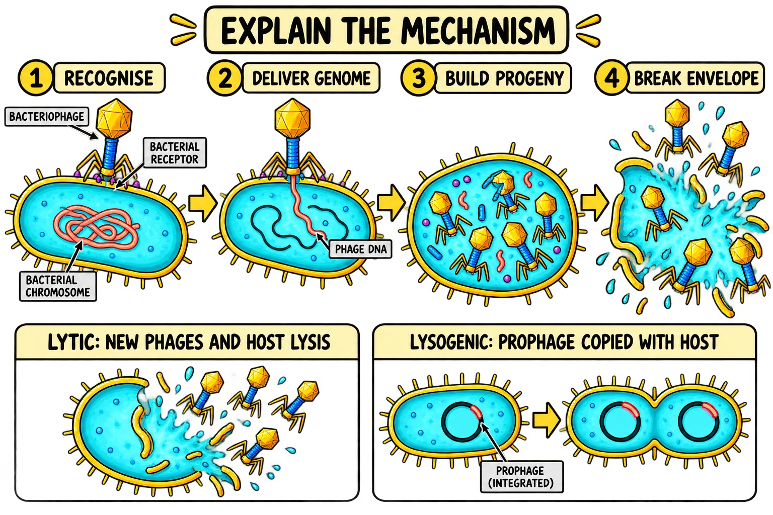
When you revise, build explanations around the movement of genetic information, the production of new components and the fate of the host envelope. These three threads connect structure, reproduction and killing without reducing the topic to a memorised picture.
Build a short answer in the right order
Begin with a suitable phage attaching to a compatible bacterial surface. State that the genome is delivered in the tailed-phage example. Explain that viral gene expression and genome replication supply the components needed to build progeny. Finish by connecting envelope disruption to release and host death in the lytic pathway. That sequence answers both “how more phages appear” and “how the bacterium is killed”.
If the question asks about lysogeny, change the middle of the explanation. The key event is maintenance of the phage genome in the host lineage, often as an integrated prophage, while the lytic programme is regulated. Then explain induction only if the question asks how the system can later move towards productive lytic development.
Reasoning problem 1: where did the infection stop?
Question: Phages attach to a bacterial strain and their DNA is detected inside, but no infectious progeny are recovered. Has receptor recognition failed?
Answer: The observations show that at least some attachment and entry occurred. The problem could be later: genome destruction, failure of expression or replication, an assembly defect, defective packaging or another barrier. To choose among these explanations, you need measurements that distinguish the stages. Do not call every unsuccessful infection a receptor problem.
Reasoning problem 2: why is there no release?
Question: A Gram-negative host contains assembled phage particles. Its peptidoglycan is damaged, but extracellular phage release is much lower than expected. What additional barrier should you consider?
Answer: Consider the outer membrane and the relevant release machinery, including spanins in a suitable system. The observation does not prove a spanin defect, but it makes the outer-membrane step a reasonable hypothesis. A strong answer separates a proposed explanation from an experimentally established diagnosis.
Reasoning problem 3: does an average describe every cell?
Question: A problem gives three million PFU added to one million bacterial cells. Does that mean each cell receives exactly three phages?
Answer: The nominal ratio is three PFU per cell, but it is an average based on the quantities supplied. Encounters and successful infections are not distributed equally by definition, and some particles may fail to reach or productively infect a cell. This is why a ratio alone cannot establish the exact number of infections in every cell.
Avoid these five common mistakes
“A phage is a bacterium.” A phage is a virus of bacteria. The host is a cell; the phage particle is not a bacterial cell.
“Every phage enters the lytic cycle immediately.” Temperate phages can establish lysogeny, and chronic filamentous infections show another relationship between production and release.
“Endolysin dissolves every envelope layer.” Its relevant target is peptidoglycan. Other barriers and proteins must be considered for the system in the question.
“Resistance means antibiotics and phages always fail together.” These are different susceptibility relationships. A bacterial strain’s response must be assessed for the particular antibiotic and phage.
“One successful patient proves a universal cure.” Clinical evidence must be interpreted by study design, comparison, sample size, disease setting and the actual outcomes measured.
Quick questions students ask
What is a bacteriophage in simple terms?
A bacteriophage is a virus that infects bacteria. It carries genetic information in a protective particle and relies on a suitable bacterial cell to make more phages. A phage is not itself a bacterium.
How do bacteriophages kill bacteria?
In a lytic infection, phage genes direct production of genomes and proteins, progeny particles assemble, and phage-associated machinery disrupts the bacterial envelope. The cell lyses and releases new phages. The precise lysis machinery differs between systems.
Do all phages kill their host immediately?
No. Temperate phages can establish lysogeny, maintaining their genome in the bacterial lineage without immediate lytic destruction. Some filamentous phages also produce and release particles from living cells through chronic infection.
What is the difference between lytic and lysogenic cycles?
The lytic pathway produces progeny and ends in host lysis in the examples discussed here. Lysogeny maintains the phage genome in the host lineage while productive lytic development is regulated. Induction can switch a functional prophage towards productive development.
Can bacteriophages infect human cells like human viruses?
Their natural reproductive hosts are bacteria. They do not carry out the same productive infection cycle in human cells as human viruses do. They can still interact with the human body, immune processes and the microbiome, so bacterial host specificity does not establish that every preparation or use is safe.
Can phages kill antibiotic-resistant bacteria?
A suitable phage may infect a bacterium that resists particular antibiotics, because the susceptibility mechanisms differ. That is not guaranteed for every bacterial isolate or phage. Phage susceptibility, delivery and clinical outcomes still need to be evaluated.
Can bacteria become resistant to phages?
Yes. Changes at the surface can prevent attachment, and intracellular systems can interrupt infection. Some resistance variants exist before phage exposure and become more common through selection. Phages can also evolve counter-defences.
What is a prophage?
A prophage is a phage genome maintained in association with the bacterial host lineage. In the familiar lambda-like example it is integrated into the bacterial chromosome. It is genetic information, not a collection of complete phage particles inside the cell.
Does a PFU count tell us the exact number of phage particles?
PFU is an infectivity measure defined by plaque formation on a particular host under particular assay conditions. It should not automatically be treated as the total number of physical particles or genome copies.
Will phage therapy replace all antibiotics?
The evidence does not support that claim. Phage products are evaluated for specific infections and may be studied alongside antibiotics. Controlled clinical comparisons are needed to establish which approaches provide useful benefits and acceptable risks.
Final self-check: Explain the lytic pathway aloud without looking at the diagram. Then explain one way a bacterium can interrupt it, one way a phage genome can persist without immediate lysis, and one reason a laboratory result does not automatically establish clinical benefit. If you can connect those answers, you understand the mechanism beyond the stage names.
Further reading
Explore the textbooks, experiments and clinical research behind these notes.
- The viral life cycle · OpenStax Microbiology
- Viral morphology and classification · OpenStax Biology 2e
- Yap and Rossmann (2014) · Structure and function of bacteriophage T4
- Ge and Wang (2024) · Structural mechanism of lambda-tail interaction with its bacterial receptor
- Large-scale genomic survey reveals strain-level phage-specificity determinants (2024)
- T4 structure and infection apparatus · Yap and Rossmann
- Hesselbach and Nakada (1975) · T7-associated regulation of host RNA polymerase
- Ellis and Delbrück (1939) · The growth of bacteriophage
- A promiscuous DNA-packaging machine from bacteriophage T4 (2011)
- T4 portal–neck–tail structures and genome positioning (2026)
- Structure and assembly of filamentous bacteriophages (2014)
- Young (2014) · Phage lysis: three steps, three choices, one outcome
- Savva et al. (2008) · Lambda holin forms rings with large diameter
- Phage lysis: multiple genes for multiple barriers (2019)
- Membrane fusion during phage lysis (2015)
- The mechanism of the phage-encoded protein antibiotic from ΦX174 (2023)
- Imaging single filamentous phages extruding from living bacteria (2013)
- Morelli et al. (2009) · DNA looping and the lambda switch
- The bacterial toxin colibactin triggers prophage induction (2022)
- Zinder and Lederberg (1952) · Genetic exchange in Salmonella
- Genetics · Medical Microbiology, NCBI Bookshelf
- Waldor and Mekalanos (1996) · A filamentous phage encoding cholera toxin
- Chan et al. (2016) · Phage selection restores antibiotic sensitivity in a studied Pseudomonas system
- Barrangou et al. (2007) · CRISPR provides acquired resistance against viruses
- Doron et al. (2018) · Systematic discovery of antiphage defence systems
- Liu et al. (2026) · Discovery of bacterial antiphage systems through protein-domain patterns
- Luria and Delbrück (1943) · Mutations from virus sensitivity to virus resistance
- Bacteriophages suppress CRISPR–Cas immunity using RNA-based anti-CRISPRs (2023)
- Regional variation in lytic and lysogenic infection in the Southern Ocean (2012)
- Hsu et al. (2019) · Phages alter a mouse-model gut microbiota and metabolome
- Barr et al. (2013) · Bacteriophage adherence to mucus
- Hyman and Abedon (2009) · Determining phage growth parameters
- Hershey and Chase (1952) · Functions of phage protein and nucleic acid
- WHO Regional Office for Europe (2025) · Building evidence for bacteriophage therapy
- TATFAR (2025) · Considerations and perspectives on phage therapy
- Dedrick et al. (2019) · Phage treatment in a patient with disseminated Mycobacterium abscessus
- Weiner et al. (2025) · BX004-A randomised first-in-human trial
- Author correction to the BX004-A paper (27 February 2026)
- FDA and NIAID · Science and regulation of bacteriophage therapy